ODT | MARCH 3, 2023. The pain came out of nowhere.
Like the flick of a switch, the ache exploded within the entrails of Asha Morris’ lower left leg and intensified by the hour. By day’s end, the Queensland teenager could no longer walk.
An MRI showed a cancerous tumor—Ewing sarcoma, actually—pressing on the leg nerve. The rare bone and soft tissue malignancy usually afflicts teenagers, though it can present in older individuals as well. “The tumour was inside the bone and into the tissue,” Morris told The Australian Women’s Weekly in December 2022, “…the soft tissue at the back of the calf behind the skin.”
Morris immediately began hormone stimulation treatment, then a year of chemotherapy that bookended tumor removal surgery. She surrendered 16 cm (6.3 in.) of bone in an experimental procedure that had only been executed once before, but not on an immunocompromised chemotherapy patient.
The technique that saved Morris’ lower leg from amputation was developed by Osteopore International Pte. Ltd., a Singapore company that designs, develops, and markets bio- resorbable polymer implants for neurosurgical, orthopedic, and maxillofacial applications. Its technology harnesses the body’s natural healing process to restore lost tissue through customized, 3D-printed bioresorbable bone implants.
Osteopore’s implants are tailor-built from patients’ computed tomography (CT) scans, and 3D printed using U.S. Food and Drug Administration (FDA)-approved polycaprolactone (PCL), a polymer with mechanical strength similar to that of porous trabecular bone. In addition to its bioresorbability, PCL is malleable and degrades slowly, making it an ideal material candidate for 3D-printed bone implants.
“We use the body’s regenerative ability to rebuild lost tissues by leveraging tissue engineering, regenerative medicine, and 3D printing techniques of biomimetic scaffolds,” Osteopore Chief Technology Officer/Chief Operating Officer Jing Lim, Ph.D., explained to Malaysia’s oldest newspaper, New Straits Times, last fall. “For example, with a 3D printer, we can make bioabsorbable implants for bones that can naturally break down into water and carbon dioxide as the healing takes place, leaving a healthy regenerated bone in its place. It also imitates the healing process to accelerate reconstruction and enable the affected bone to heal on its own.”
Osteopore’s implants completely break down in 18 to 24 months’ time. Their honeycomb microstructure and interconnected pores, according to the company, support blood vessel and new bone growth both on the surface and deep within the material, thus enabling the body to regenerate lost bone. New tissue, however, is regenerated only in the bone defect and the entire process lasts only as long as necessary.
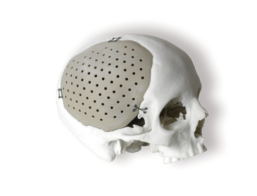
Studies have shown Osteopore’s implants foster vascularization by creating a direct mineralized matrix within the scaffold interior. Backed by more than a decade of clinical data, the products have been used in over 60,000 successful procedures globally to regenerate bones in the forearm, shoulder, skull, jaw, and face; nasal reconstruction is forthcoming as the company works to expand its portfolio.
Currently cleared in the United States, Europe, Australia, and Singapore (China’s in the works), and distributed in more than 20 countries worldwide, Osteopore implants purportedly have a flawless performance record. The company touts the absence of any adverse or serious events, and claims its products are less likely than traditional metal/plastic bone replacement solutions to cause infections or post-operative complications. Such superb performance metrics result in lower overall medical costs (fewer, if any, follow-up surgeries) and better patient outcomes, Osteopore executives boast.
Morris certainly can attest to those improved patient outcomes, as she was more affected by chemotherapy than the implant surgery. Besides robbing the girl of her hair, muscle mass, and a significant amount of weight, the cell-killing cancer treatment also poached her overall immunity—triggering multiple infections—and her healthy mucosal tissue, causing a nasty bout of mucositis.
There was, however, one thing cancer couldn’t steal from Morris—a positive attitude. That optimism was as integral to her recovery as the chemotherapy and implant surgery. Even now, 29 months post-diagnosis, Morris refuses to reflect upon her ordeal in a negative manner.
Quite the contrary, in fact.
“…with something bad comes something good. Your cancer isn’t your identity, it’s a part of your puzzle and that pathway or experience could be a part of something down the track,” she noted to AWW. “…having cancer has been the greatest blessing to me because I’m so appreciative of everything, and I don’t think that will go away. To be 19 and truly live with intention and purpose and awareness of just being alive and make [a] connection with people and walk on my own legs—these are things I took for granted. I feel blessed to be able to live healthily. I feel very grateful for the gifts that my cancer gave me.”
Indeed, Morris is blessed to be living healthfully now, but she’s equally as blessed to be living in an age of such advanced, science-fiction-like technologies as in-situ tissue engineering and 3D printing, both of which were still in their infancies a mere 10 years ago.
Though its origins can be traced to a 1971 liquid metal extruder patent filing (courtesy of inventor Johannes F. Gottwald), 3D printing technology evolved slowly at first, as the stereolithography process required decades of research and fine-tuning to make it commercially viable. The first machines were large, slow, and costly (upwards of $300,000), and still needed some amount of manual intervention.
By the turn of the millennium, 3D printing technology had advanced enough to produce dental implants and custom prosthetics—applications that surprised stereolithography originator Charles W. Hall, who never expected the manufacturing technique to be used in medicine.
Further developments over the next dozen years or so spanned science-fiction-like innovations—i.e., a fully functioning miniature kidney, a bladder, blood vessels, tissue cartilage, and a lower jaw that was implanted into an 83-year-old woman suffering from a chronic bone infection.
3D printing has become a particularly useful tool in orthopedics. Not only has the technology helped improve pre-operative planning, it’s also enabled surgeons to create patient- specific instruments and customized implants and devices.
Such customization can be found in the 3D printed titanium components made by Stryker Corp. and the Cleveland Clinic; the Vault shoulder reconstruction system and triflange hip replacement products from Zimmer Biomet Holdings Inc.; and the bio-sourced, recyclable orthosis launched last spring by digital manufacturing provider Sculpteo and Daniel Robert Orthopedic.
Customization also manifests itself in Conformis’s 3D printed iTotal joint replacement system, which generates tailor-made instrumentation called iJigs for personalized hip and knee replacements. In addition, the Billerica, Mass.-based firm uses 3D printing (a.k.a., additive manufacturing) to make the wax molds in which its metal implants are cast.
Patient specificity is the goal as well for engineers at the University of Bath’s Centre for Therapeutic Innovation in the United Kingdom, who have developed a new treatment for knee osteoarthritis. Dubbed Tailored Osteotomy for Knee Alignment (TOKA), the treatment uses 3D printing to improve the operative procedure and fit of high-tibial osteotomy (HTO) plates for knee realignment. Currently in clinical trials, the personalized, titanium-alloy plates are said to be more stable, comfortable, and better able to bear weight than existing generic plates.1 They also shorten HTO surgical time, thus improving the safety of such procedures, according to the University of Bath engineers.
Optimally positioned and secured against the bone using 3D printed screw threads (an industry first), the plates have been safety tested virtually in a computer-based trial using CT scan data from 28 patients.1
“Additive manufacturing (AM) continues to gain widespread adoption in the orthopedic industry,” said Gaurav Lalwani, Ph.D., Global Applications Engineering lead for Medical Markets at Carpenter Technology Corporation, a Philadelphia-headquartered developer of high-performance specialty alloy-based materials and process solutions. “Medical device components such as spinal cages, acetabular cups for total hip arthroplasty and tibial base plates for total knee arthroplasty are routinely printed using electron beam and laser powder bed fusion processes. Emerging trends such as printing patient-specific surgical guides and instrumentation have been explored wherein OEMs don’t need to invest in specialized tooling and subtractive manufacturing processes for low-rate customized production runs.”
Patient-specific surgical guides and instruments are key elements of most 3D printed orthopedic solutions. Crafted from CT images of patient anatomy, the guides help simplify the pre-operative planning process and boost overall procedure success rates by eliminating the possibility of excess bone removal or inaccurate component placement.
Software packages such as Materialize 3-matic (Materialise NV), Windows 3D Builder (Microsoft Corporation), Rhino (Robert McNeel & Associates), ThinkerCAD and Meshmixer (both Autodesk) enable surgeons to plan specific procedures, determine osteotomy locations or planes, or simulate resections. Synopsys’s Scan IP software module, Simpleware AS Ortho, shortens segmentation times and quickly produces accurate results via automated algorithms. Specifically designed for hip and knee segmentation purposes, ScanIP software generates a 20 to 50 times faster segmentation rate for clinical images, the company boasts.
“In software, there have been advancements in developing algorithms to help the segmentation phase, accelerating the creation of 3D models from diagnostic images. Software has also been developed to help design complex shapes to accommodate personalized anatomy,” noted Francesco Robotti, technology business development manager at Lincotek Medical, a global contract manufacturer that provides casting, machining, plasma spray coatings, and other technologies for the medical industry. “Other software is helping design porous-lattice structures, simplifying the computational work for the benefits of faster design and faster printing execution.”
That kind of software currently is offered by global SaaS firm Oqton. Its 3DXpert is touted as an “all-in-one integrated software” that streamlines the 3D printing process by optimizing component design structure, shortening the design to production lead time, and minimizing the manufacturing total cost of operation.
3DXpert can produce hollowed out parts that maintain their shape and meet mechanical specifications, according to the company. Its vast library of pre-defined lattice structures serves as the basis for original lattice designs or those modeled after other systems. The software also allows engineers to run an FEA stress analysis on lattice structure and optimize lattice elements based on that evaluation to meet functional properties requirements while minimizing weight, material usage, and printing time.
“Oqton’s 3DXpert software utilizes sophisticated support strategies that minimize the post-processing of the printed parts, thereby reducing the overall cost of the part,” explained Gautam Gupta, Ph.D., senior vice president and general manager, Medical Devices, at 3D Systems, a Rock Hill, S.C.-based additive manufacturing solutions partner. “In addition, 3DXpert has unmatched credibility, as there are dozens of FDA-cleared and CE-marked devices that utilize the software to integrate complex bone ingrowth porous structures to improve the overall performance of the device.”
Machine Multiplicity
Most 3D printed plastics are created through stereolithography (SLA), selective laser sintering (SLS), or fused deposition modeling (FDM). SLA printers cure liquid resin layer by layer through ultraviolet laser light, generating precise, high -resolution objects with material versatility.
SLS printers use high-powered lasers to sinter (bind) powdered material into a solid structure. The machine lays down an even powder layer, sinters that layer, then repeats the process until the part is complete. This printing technique shapes components by aiming a laser at the powder bed in specific points in space using a digitally produced computer-aided design file as a guide. SLS’s vast material range makes it an ideal choice for complex mechanical parts.
FDM printing, also known as fused filament fabrication, is most commonly used for rapid prototype production, though it is also well-suited for tissue engineering and patient-specific prosthetic and bone implants. Patented by AM machine developer Stratasys Ltd., FDM builds parts in layers by selectively depositing melted material in a pre-determined path, using thermoplastic polymer filaments to form the final object.
The company’s J850 Digital Anatomy 3D printer creates three-dimensional models of human anatomy that mimic bone and tissue. It can create regions for screw entry, enabling clinicians to place screws without cracking the model, and autogenerate all bone region structures—proximal, distal, cortical, cancellous, and the medullary canal.
Stratasys’s J5 MediJet 3D Printer’s multi-material and multicolor capabilities produce vivid anatomical models as well as drilling and cutting guides that are sterilizable and biocompatible, with a certified system.
The Kumovis R1 printer from Munich-based Kumovis GmbH, which was acquired by 3D Systems last year, creates devices from implantable medical-grade polymers like polyetheretherketone (PEEK) and Radel PPSU (polyphenylsulfone). It reportedly is the only extrusion plat- form with a build chamber that can be turned into a clinically validated cleanroom. Designed to control temperatures during production, the printer is equipped with a global laminar air flow, which heats the build chamber homogenously up to 250 degrees Celsius. R1 users can adjust local cooling for each individual strand and layer, heat up to further increase layer adhesion, reduce the need for post-processing, and simplify support structure removal.
Troy, N.Y.-based Lithoz has developed a printer designed specifically for customizing oxide ceramics. Launched in April 2021, the CeraFab Lab L30 features 50 µm resolution and uses the company’s ceramic slurry-based technology, which dips a build platform in the slurry and then illuminates it from beneath using LED. Parts are subsequently debound and sintered.
The EOS M 290, M 300 Series, M 400, and M 400-4 printers lack Lithoz’s ceramics prowess or Kumovis’ build chamber-cleanroom conversion but they have extensive material portfolios and can achieve densities at nearly 100%. The direct metal laser sintering machines also can produce lattice and honeycomb designs for optimal osteoconductive behavior.
Direct metal laser sintering (DMLS) and selective laser melting (SLM) printers work similarly to SLS machines but fuse metal powder particles together by layer with a laser. Both kinds of printers create strong, complex metal products commonly used in orthopedic applications.
Earlier this year, contract manufacturer rms Company added 3D Systems’s DMP Flex 350 Dual printer to its production workflow in order to expand its implants offering. The DMP Flex 350 Dual is the most recent addition to 3D Systems’s Direct Metal Printing portfolio. Its dual-laser configuration maintains the benefits of the single-laser configuration, including flexible application use and quick-swap build modules, as well as a central server to manage print jobs, materials, settings, and maintenance for 24/7 productivity. Additionally, the company’s unique vacuum chamber significantly reduces argon gas consumption while delivering best-in-class oxygen purity (<25 ppm). The printer also is equipped with Oqton’s 3DXpert software.
“In metals, we are focusing our innovation efforts on increasing the productivity of our systems and reducing the final part cost for customers,” Gupta told ODT. “Toward achieving that goal, we have launched the DMP Flex 350 Dual, a dual-laser system that can print in 90-micron layers due to high-wattage lasers while maintaining mechanical properties and surface resolution, and accuracy. Because we can do this in both Titanium and CoCr alloys, it generates a lot of interest within the industry for applications in total joint replacement.”
‘Smarter’ Materials?
Self-sufficient and “smart.”
Those adjectives reflect the future potential of 3D-printed orthopedic implants.
That potential is currently underway at the University of Pittsburgh Swanson School of Engineering, where researchers are developing patient-specific 3D printed “smart” metamaterial implants that double as sensors to monitor spinal healing. The school’s Intelligent Structural Monitoring and Response Testing (iSMaRT) Lab has created a new class of multifunctional mechanical metamaterials that act as their own sensors, recording and relaying data about structural pressure and stresses. These self-aware metamaterialas, other- wise known as “meta tribomaterials,’ generate their own power and can be used for various sensing and monitoring applications.
The material is designed to produce contact-electrification under pressure between its conductive and dielectic microlayers, which creates an electric charge that relays information about the material matrix’s condition. In addition, it naturally inherits the mechanical tunability of standard metamaterials. The power generated from its built-in triboelectric nanogenerator mechanism eliminates the need for a separate power source, and a tiny chip records data about cage pressure, an important healing indicator. The data can then be read noninvasively with a portable ultrasound scanner.
Not only is the proposed cage unique in its sensing capabilities, it’s also made of a highly tunable material that can be customized to the patient’s needs.
“This is a first-of-its-kind implant that leverages advances in nanogenerators and metamaterial to build multifunctionality into the fabric of medical implants,” said Amir Alavi, assistant professor of civil and environmental engineering, whose iSMaRT Lab led the research. “This technological advancement is going to play a major part in the future of implantable devices.”
Until that future actually arrives, however, 3D printing mavens have metals like titanium and cobalt chrome and polymers like PEEK at their disposal. Research has shown the porous structure of 3D printed titanium to be comparable to skeletal bones; the lattice structures’ rough texture promotes osseointegration and facilitates soft tissue and bone regrowth via nutrients that flow around the structure.
LimaCorporate S.p.A. and the Hospital for Special Surgery (HSS), partnered several years ago to establish the first AM facility for complex custom implants in a hospital setting, using Lima’s patented 3D printed Trabecular Titanium implants for patient-specific solutions.
“Materials development in the 3D printing space takes time, particularly for medical. Metals are currently widely used, including titanium and 17-4 Stainless Steel, primarily,” said Brian McLaughlin, president of ARCH Additive, a division of precision machining, contract manufacturing, and supply chain integration resource firm ARCH Medical Solutions. “Other materials used for medical devices that can be printed include CoCr and 316SS. Work continues on biologic scaffolds with the idea of incorporating stems cells and or similar means for tissue differentiation. Ceramics are also being worked on…”
The ceramics under development mostly comprise alumina and zirconia—high-density materials with good mechanical strength and corrosion resistance—and hydroxyapatite, a calcium phosphate found in teeth and bone. Lithoz has improved upon the latter bioceramic in its new bone graft substitute, LithaBone HA 480. Unveiled late last year, LithaBone increases the wall thickness (from <1.6mm to 10mm) and further reduces minimum wall thickness, broadening the range of its applications.
LithaBone HA 480 also has less overpolymerization but an improved depth of cure, which improves build parameters and induces a more stable manufacturing process. The material also has a longer shelf life and produces easier-to-clean parts.
SINTEX Technologies Inc.’s acquisition last summer of Technology Assessment and Transfer Inc. provided the Salt Lake City-based firm with the necessary processing equipment and novel composite filaments for manufacturing 3D printed implants from its silicone nitride material. SINTEX’s FleX-SN product family can be polished to a smooth and wear-resistant surface for articulating applications, such as bearings for hip and knee replacements. They are suitable as a dental implant material as well, thanks to their bacterial adhesion resistance.
3D Systems made a similar move last spring to diversify its AM capabilities from metal implants to PEEK products. Company executives said the purchase of Kumovis would benefit 3D Systems’s portfolio of craniomaxillofacial, bone plate, and spinal cage applications.
While AM has grown significantly in the orthopedic industry over the last two decades, experts say there are still some large-scale hurdles to overcome before the technology is used to its full advantage—cost, hardware, software, materials, and services, to name a few.
“We believe cost is still a challenge for 3D printing of components that are relatively large, driving low yields on a print run, and where markets are price sensitive, such as hip or knee replacement,” said Dave Anderson, co-founder and business development leader at Mach Medical, a Columbia City, Ind.-based orthopedic medical device contract manufacturer. “Adding to the cost challenge is the fact that, in many applications, 3D printing forces additional steps to the manufacturing process. For instance, 3D printed products will often require printing, heat treatments, cleaning, and inspection as incremental steps to the traditional process. This can add lead time, which will unfavorably impact inventory levels, and cost, which is harder to absorb with product price.”
Juan Chafino added regulatory validations and AM’s part-dependent nature to the list of hurdles the technology still faces. But he’s hopeful for the future, as he believes the technique is still in its infancy.
“As the technology matures, the process and process flow will become more industrial. As the market learns the new design rules and think out of the box, we will see that medical devices are more suited for 3D printing,” noted Chafino, additive manufacturing expert at orthopedic instrument and implant developer/manufacturer InTech, Chatellerault, France. “The future of 3D printing is linked to the advances on software technologies in terms of applications and technology itself. An orthopedic digital twin of a human body will unlock the full potential of patient-specific implants for simulation purposes to ensure the kinematics are optional, which will improve patient outcomes while extending the lifespan of the medical device itself.”
References